Skin Longevity | 3D Skin Architecture Profiling by Dr. Amr Ismail

3D Skin Architecture Profiling
Evaluate your cellular aging timeline across the three clinical vectors: The Spark, The Brain, and The Bone.
SKIN LONGEVITY | 3D SKIN ARCHITECTURE PROFILING | Dr. Amr Ismail, MD
Direct Monograph Acquisition Hub
Bypass the profiling engine and secure immediate, uncompromised access to independent medical biology protocols.
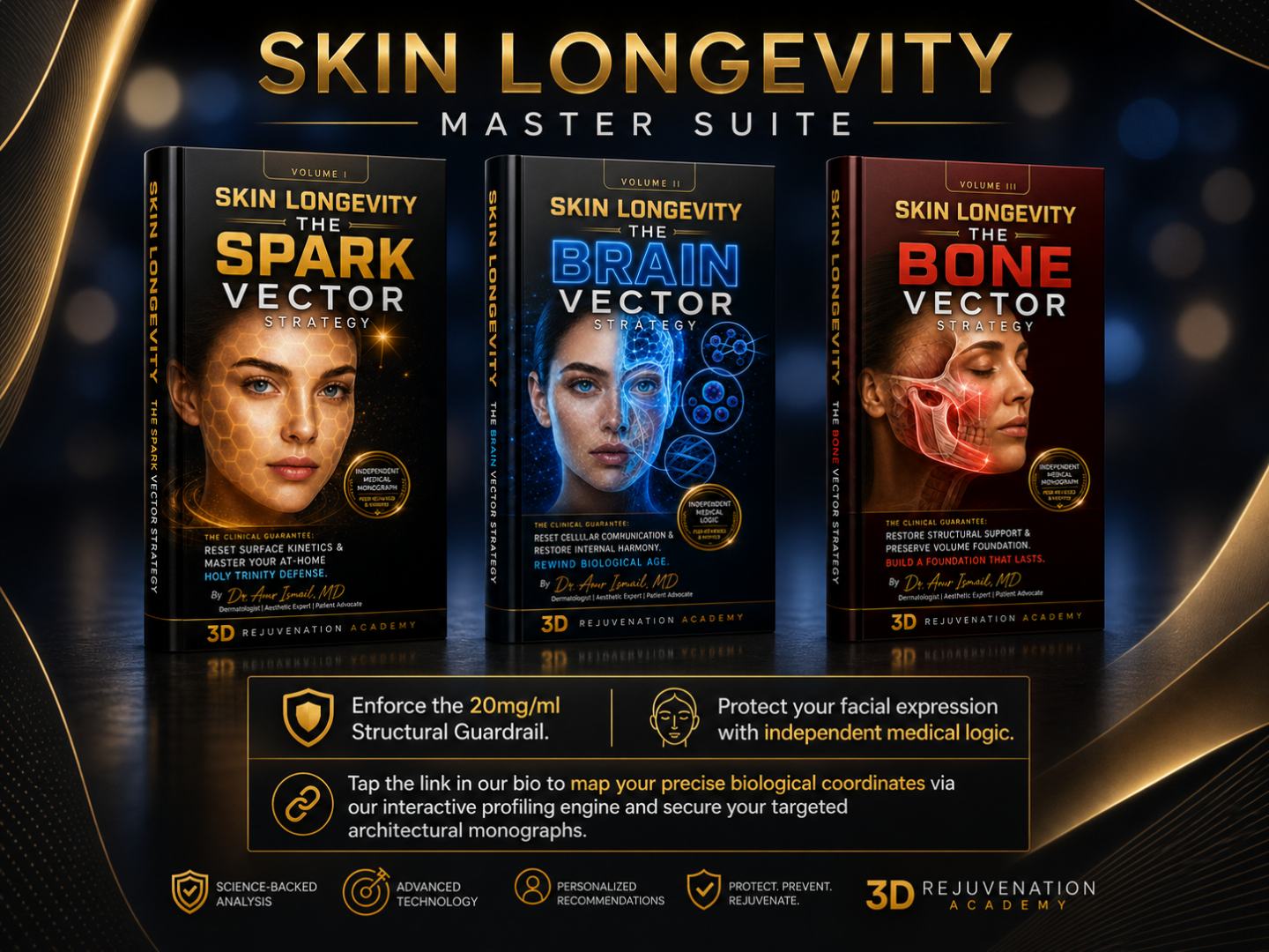
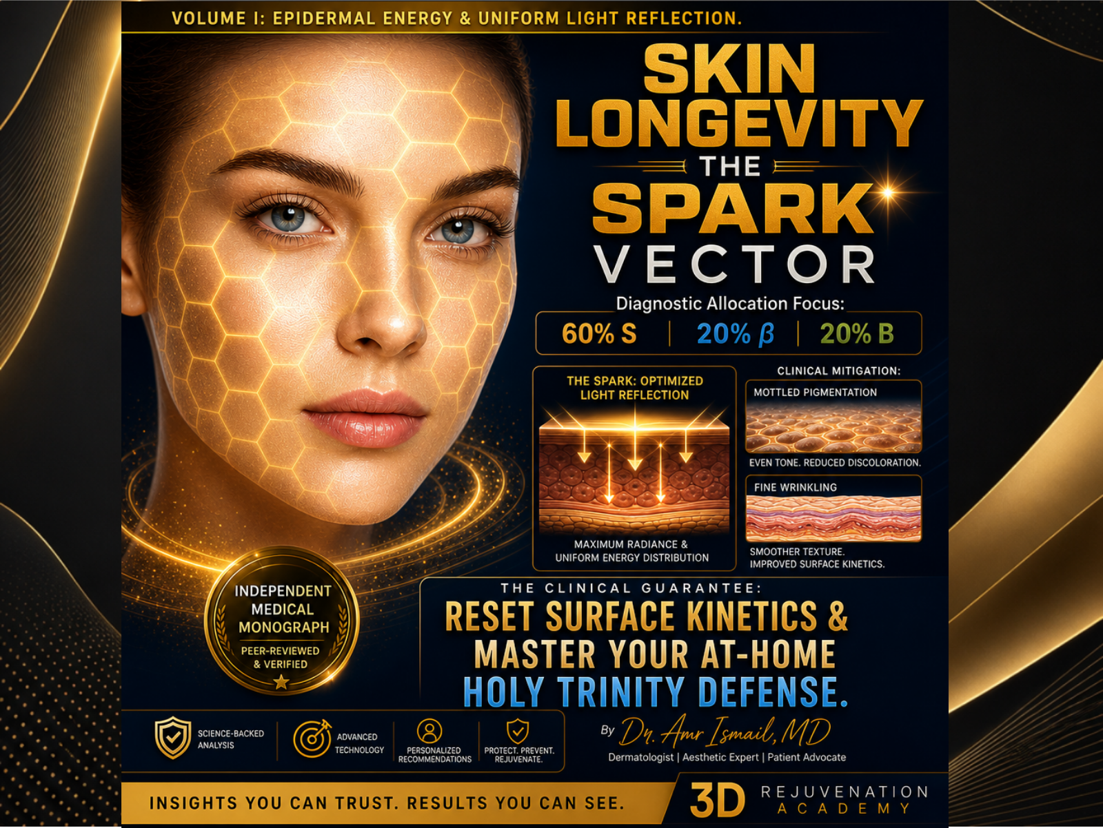
Reset your skin’s optical surface mirror, accelerate basal layer mitosis, and master the at-home Holy Trinity formulation parameters.
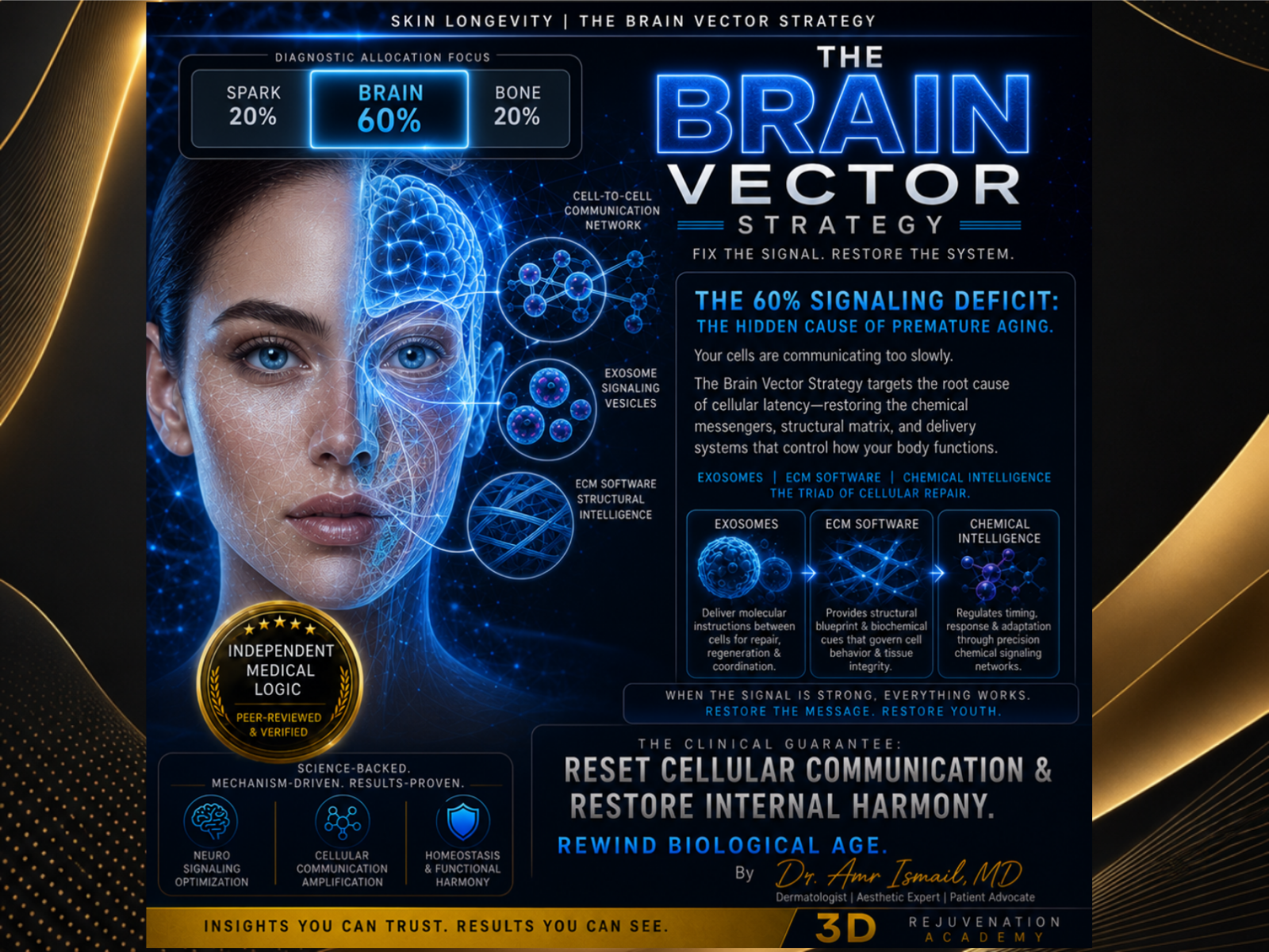
Debug corrupted ECM software loops. Deploy the advanced chemical intelligence of stem-cell-derived Exosomes and PDRN Liquid Gold.
Rebuild deep tissue vertical suspension anchors without added artificial volume. Enforce the strict 20mg/ml concentration guardrail.
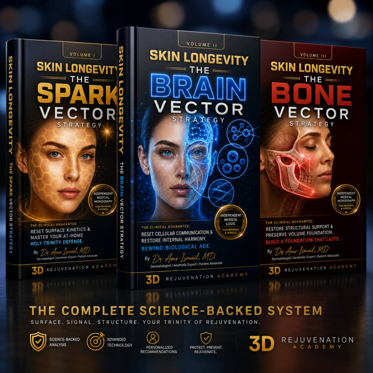
The ultimate architectural reset. Combines the Spark, Brain, and Bone blueprints into a singular, uncompromised, high-yield digital library.
Ligamentous Laxity vs. Artificial Volume: Why Over-Injecting Hyaluronic Acid Destroys Natural Facial Expressions
When a mirror reveals the early descent of the lower face—manifesting as a softening jawline, pronounced tear troughs, or the structural folding of jowls—modern cosmetic marketing presents a singular, simplistic answer: volume.
We are told that aging is a simple math problem of volume depletion, and that the immediate solution is the structural placement of cross-linked hyaluronic acid gels.
This is a profound anatomical misunderstanding.
True structural descent is rarely a failure of superficial volume; it is a failure of tensile architecture.
When clinical injectors try to lift sagging, lax tissue by pumping heavy, moisture-binding gels into the superficial layers of the skin, they do not restore youth. They simply stretch the cutaneous envelope.
The result is the distortion of natural facial physics—an over-filled, immobile presentation often referred to as "pillow face".
To reverse descent without sacrificing the biological intelligence of your natural expressions, you must stop treating your face like a balloon to be inflated and start treating it like an architectural masterpiece to be scaffolded.
🏛️ The Biomechanical Reality: Ligamentous Laxity
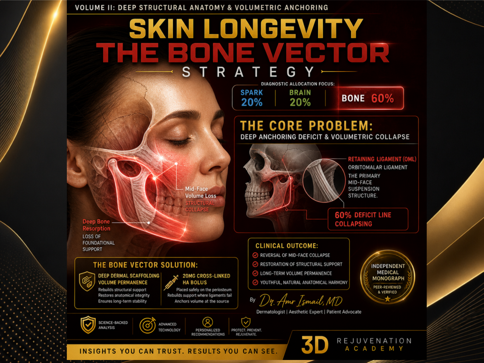
Your facial soft tissues are anchored to the underlying facial skeleton by a network of dense, fibrous bands called retaining ligaments.
In the midface and periorbital regions, the two most critical structural anchors are:
- The Orbitomalar Ligament (OML): Anchors the lower eyelid tissue to the bony orbital rim.
- The Zygomatic Cutaneous Ligament (ZCL): Anchors the cheek fat pads directly to the zygomatic bone.
Think of these ligaments as vertical suspension cables. In your 20s and 30s, these cables are tight, holding fat pads and deep tissue layers firmly against the skeletal frame.
As the biological timeline advances, these fibrous bands experience structural elongation. The cables stretch.
[ Young Architecture ]          [ Advanced Laxity ]
     periosteum                      periosteum
        │                               │
   (Tight Cable)                  (Stretched Cable)
        ▼                               ▼
 [ Firm Soft Tissue ]           [ Sagging Soft Tissue ] (Jowls / Bags)
When these ligaments stretch, the soft tissue slides downward over the bony anatomy, pooling along the jawline to create jowls, or separating under the eye to create deep tear troughs. The underlying bone itself undergoes slow resorption, reducing the projecting shelf that these ligaments rely on for leverage.
🛑 The Industry Failure: The Superficial Hydration Trap
When a stretched structural ligament causes tissue to sag, the skin above it folds. The commercial cosmetic industry looks at this fold and attempts to "fill the wrinkle".
Hyaluronic acid (HA) dermal fillers are highly hydrophilic—they are designed to bind up to 1,000 times their weight in water. When an injector places a large bolus of this water-loving gel into the superficial fat pads of a sagging cheek, the gel absorbs local water metrics and swells.
This creates a brief optical illusion of a lift by strictly stretching the skin taut. However, it introduces a severe mechanical problem: gravitational weight.
- Heavy, water-logged gels add physical mass to an area that is already sliding downward due to weak ligaments.
- Over a multi-month timeline, this added weight accelerates the elongation of the Zygomatic Cutaneous Ligament, dragging the midface down even faster.
- When you smile, your natural facial muscles (like the zygosomaticus major) attempt to lift this heavy, artificial mass. Because the gel cannot compress or mimic real fat tissue, it pushes upward and outward as a stiff, unnatural block.
This is what erases your natural expressions, creating a puffy, swollen appearance that looks entirely foreign to your biological identity. In thin periorbital zones, superficial placement leads to water pooling, chronic edema, and the blue hue of the Tyndall effect.
📐 The Architectural Reset: Deep Structural Scaffolding
To restore lower-face tension and lift sagging jowls without distorting your expressions, you must shift your clinical parameters entirely away from superficial soft-tissue filling. You must deploy the principles of The Bone Vector.
True structural age-reversal requires deep, supraperiosteal bolus placement. Instead of placing filler into the skin, highly cohesive, high-G* prime gels must be placed deeply underneath the muscle layers, directly onto the bone.
By placing small, precise droplets of product right against the periosteum at the true ligamentous insertion points, you create a rigid mechanical strut. This deep strut changes the angle of the stretched ligament, pulling the cable taut from underneath and restoring its vertical suspension capabilities.
[ SUPRAPERIOSTEAL REFLECTION ]
  [ Deep Bone Frame ] ➔ [ High G* Cohesive Strut ] ➔ [ Tightened Structural Ligament ] ➔ [ Natural Lift ]
This approach adds no volume to the moving, expressive layers of your face. Your superficial fat pads remain light and free to move naturally with your facial muscles, completely protecting your genuine expressions.
🛡️ Enforcing the 20mg/ml Structural Guardrail
When utilizing deep supraperiosteal placement, you must protect your tissues from chronic, low-grade inflammatory swelling. The thinner the tissue zone, the more rigid your safety boundaries must be.
In delicate zones like the periorbital matrix or the midface tear trough transition, you must strictly enforce a 20mg/ml Concentration Guardrail.
High-density hyaluronic acid gels exceeding a 20mg/ml concentration possess excessive swelling pressures. They act as constant water magnets, distorting the fine contours of your lower eyelid and midface.
Demand that any intervention in these zones utilizes a highly cohesive, low-concentration formulation (at or below 20mg/ml) that prioritizes structural shape retention over water absorption.
🎓 Secure Your Uncompromised Architectural Protocol
Stop allowing commercial marketing to treat your facial anatomy like a hollow vessel to be over-inflated. Real age restoration is a discipline of vertical tension, skeletal support, and uncompromised structural rules.
Bypass the superficial noise of retail beauty advice and master the exact biological blueprints of your facial layers.
- Are you experiencing profound midface descent or tear trough hollowing? Secure immediate, step-by-step guidance on structural safety boundaries by downloading Volume III: The Bone Vector Monograph directly from the 3D Rejuvenation Academy.
- Ready to master the entire framework? Protect your surface, signaling, and deep skeletal architecture simultaneously by acquiring the unified 3-Volume Collection Suite Bundle at a specialized tier today.